
Diabetic Eye Exams: Screening & Diagnosis
Diabetic eye disease can develop before you notice any changes in your vision. A diabetic eye examination allows an eye-care professional to look for changes in the eye so that problems can be identified and managed as early as possible.
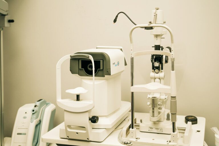
Why Are Diabetic Eye Exams Important?
Diabetic retinopathy often has no symptoms in its early stages. Even people with more advanced disease may sometimes have little or no noticeable change in vision.
An eye examination can identify retinal changes before they begin affecting everyday vision. This gives your healthcare team an opportunity to monitor the condition or consider treatment when appropriate.
A diabetic eye examination can also identify other eye conditions that may occur more often in people with diabetes, including cataracts and glaucoma.
When Should People With Diabetes Have Their Eyes Examined?
Screening recommendations depend on the type of diabetes and the individual’s eye health.
For adults with type 1 diabetes, the American Diabetes Association’s 2026 Standards of Care recommend an initial comprehensive dilated eye examination approximately five years after the onset of diabetes.
For adults with type 2 diabetes, the initial comprehensive dilated eye examination is recommended at the time of diabetes diagnosis because retinal changes may already be present when type 2 diabetes is first identified.
After the initial examination, the appropriate interval depends on the findings. If there is no evidence of retinopathy and diabetes is well controlled, examinations every one to two years may be considered in some patients. If diabetic retinopathy is present, examinations are generally needed at least annually and may be needed more frequently when disease is progressing or sight-threatening.
People with pre-existing type 1 or type 2 diabetes who are planning pregnancy or become pregnant may need additional examinations because pregnancy can affect the risk of diabetic retinopathy progression.
Your eye-care professional should determine the schedule that is appropriate for you.

What Happens During a Diabetic Eye Examination?
A diabetic eye examination usually begins with a discussion about your medical and eye history.
Initial Inquiry
Your eye-care professional may ask about:
- The type of diabetes you have
- How long you have had diabetes
- Changes in your vision
- Previous eye conditions or treatments
- Medications
- Blood sugar or A1C management
- Blood pressure and cholesterol
- Pregnancy or plans for pregnancy when relevant
The examination may include several different tests.
Dilated Eye Examination
A dilated eye examination is one of the most important parts of diabetic eye screening.
Eye drops are used to widen the pupils. This allows the eye-care professional to see more of the retina and look for changes such as damaged or leaking blood vessels, bleeding, retinal swelling, and abnormal blood vessel growth.
The examination itself is generally painless. The dilating drops can temporarily make your vision blurry and increase sensitivity to light, so it may be helpful to plan for temporary changes in your vision after the appointment.
Visual Acuity Testing
Visual acuity testing measures how clearly you can see at different distances.
It is useful for documenting your current level of vision and identifying changes over time. However, normal visual acuity does not rule out diabetic retinopathy because retinal damage can exist before noticeable vision loss occurs.
Retinal Examination
After the pupils are dilated, the eye-care professional examines the retina for signs of diabetic damage.
They may look for small areas of blood-vessel leakage, bleeding, retinal swelling, abnormal blood vessels, or other changes associated with diabetic retinopathy.
The findings help determine whether there is no retinopathy, an early stage of disease, or more advanced disease requiring closer monitoring or treatment.
Retinal Photography
Retinal photographs provide images of the back of the eye that can be reviewed and compared over time.
Retinal photography can be useful for diabetic retinopathy screening, including certain remote or technology-assisted screening programs. However, abnormal or inadequate images require an appropriate pathway for comprehensive eye evaluation, and retinal photography does not replace a comprehensive dilated examination in every situation.
Optical Coherence Tomography
An optical coherence tomography, or OCT, scan creates detailed images of the retina.
It can help an eye-care professional identify and measure retinal swelling, including swelling associated with diabetic macular edema. The test is noninvasive and does not require an injection.
OCT may be especially useful when a doctor needs more information about the macula or wants to monitor changes over time.
Fluorescein Angiography
If more advanced diabetic retinopathy or diabetic macular edema is suspected, an eye-care professional may recommend fluorescein angiography.
During this test, a special dye is injected into a vein, usually in the arm. A specialized camera then takes photographs as the dye travels through the blood vessels of the retina.
This can help identify areas where retinal blood vessels are leaking or where blood flow is abnormal.
Not every person with diabetes needs fluorescein angiography.

How Is Diabetic Retinopathy Diagnosed?
The diagnosis is based on the findings from the eye examination and any additional testing that is appropriate.
Your eye-care professional may determine whether there is:
- No evidence of diabetic retinopathy
- Mild or moderate nonproliferative diabetic retinopathy
- Severe nonproliferative diabetic retinopathy
- Proliferative diabetic retinopathy
- Diabetic macular edema
- Another eye condition affecting vision
The stage and location of retinal changes help determine whether monitoring or treatment is appropriate.
What Happens If Diabetic Retinopathy Is Found?
inding diabetic retinopathy does not automatically mean that you need an immediate procedure.
Some early cases may be monitored with follow-up examinations. More advanced or vision-threatening disease may require treatment or referral to an ophthalmologist with experience managing diabetic retinal disease.
The recommended follow-up interval depends on the severity and progression of the disease.


What If My Examination Is Normal?
A normal examination is reassuring, but it does not necessarily mean that future examinations are unnecessary.
Diabetes-related retinal changes can develop over time. Your future screening schedule should be based on your type of diabetes, previous examination results, diabetes management, and other risk factors.
The American Diabetes Association notes that some people with consistently normal examinations and well-controlled diabetes may be able to extend screening intervals, while people with retinopathy generally require at least annual examinations and sometimes more frequent monitoring.
When Should You Seek Prompt Eye Care?
Do not wait for your next routine appointment if you experience a significant or sudden change in vision.
Contact an eye-care professional promptly if you develop:
- A sudden increase in floaters or dark spots
- New flashes of light
- Sudden or significant vision loss
- A dark curtain or shadow across part of your vision
- New or rapidly worsening distortion of central vision
These symptoms can occur for several reasons, including bleeding or retinal complications, and need appropriate evaluation.

Frequently Asked Questions
Is a diabetic eye exam painful?
The examination itself is generally painless. Dilating drops may cause temporary light sensitivity and blurred near vision.
Do I need a diabetic eye exam if I can see perfectly?
Yes. Diabetic retinopathy can be present before symptoms appear, so normal vision does not rule out retinal disease.
How often should I have a diabetic eye exam?
There is no single schedule that applies to everyone. The interval depends on your type of diabetes, retinal findings, blood sugar control, pregnancy status, and other factors.
Does an eye exam only check for diabetic retinopathy?
No. A comprehensive eye examination can also identify other eye conditions, including cataracts, glaucoma, and other causes of vision changes.
What is an OCT scan?
OCT is an imaging test that produces detailed pictures of the retina. It can help identify and monitor swelling associated with diabetic macular edema.
Does everyone with diabetic retinopathy need treatment?
No. Some people with earlier stages may be monitored closely, while treatment may be recommended for more advanced or vision-threatening disease.
Can a retinal photograph replace an eye examination?
Retinal photography can be useful for screening in appropriate programs, but abnormal results or inadequate images require appropriate follow-up. Comprehensive dilated examinations remain important, particularly when disease is suspected or known.
Continue Learning
Understanding Diabetic Eye Disease
Learn how diabetes affects the retina and other parts of the eye, including symptoms, risk factors, and potential complications.
Diabetic Eye Disease Treatment & Long-Term Care
Learn about monitoring, injections, laser treatment, surgery, follow-up care, and protecting vision over time.

Medical Disclaimer
The information on this page is provided for general educational purposes and is not a substitute for a professional eye examination, diagnosis, or individualized medical advice. Cataract treatment and surgical decisions depend on each patient’s eye health, medical history, visual needs, and other individual factors. If you have sudden or significant changes in vision, severe eye pain, or other concerning symptoms, seek prompt medical attention.

Ready to See the World More Clearly?
Schedule your comprehensive eye exam with Hillside Ophthalmology today and experience expert care tailored to your needs.